
₹ 8,499 ₹ 22,445

₹ 6,799 ₹ 17,659

₹ 33,899 ₹ 69,579

₹ 529 ₹ 1,394
 - MYASKRO")
₹ 25,999 ₹ 40,119
")
₹ 5,999 ₹ 14,801

₹ 6,800 ₹ 15,157

₹ 5,999 ₹ 10,319
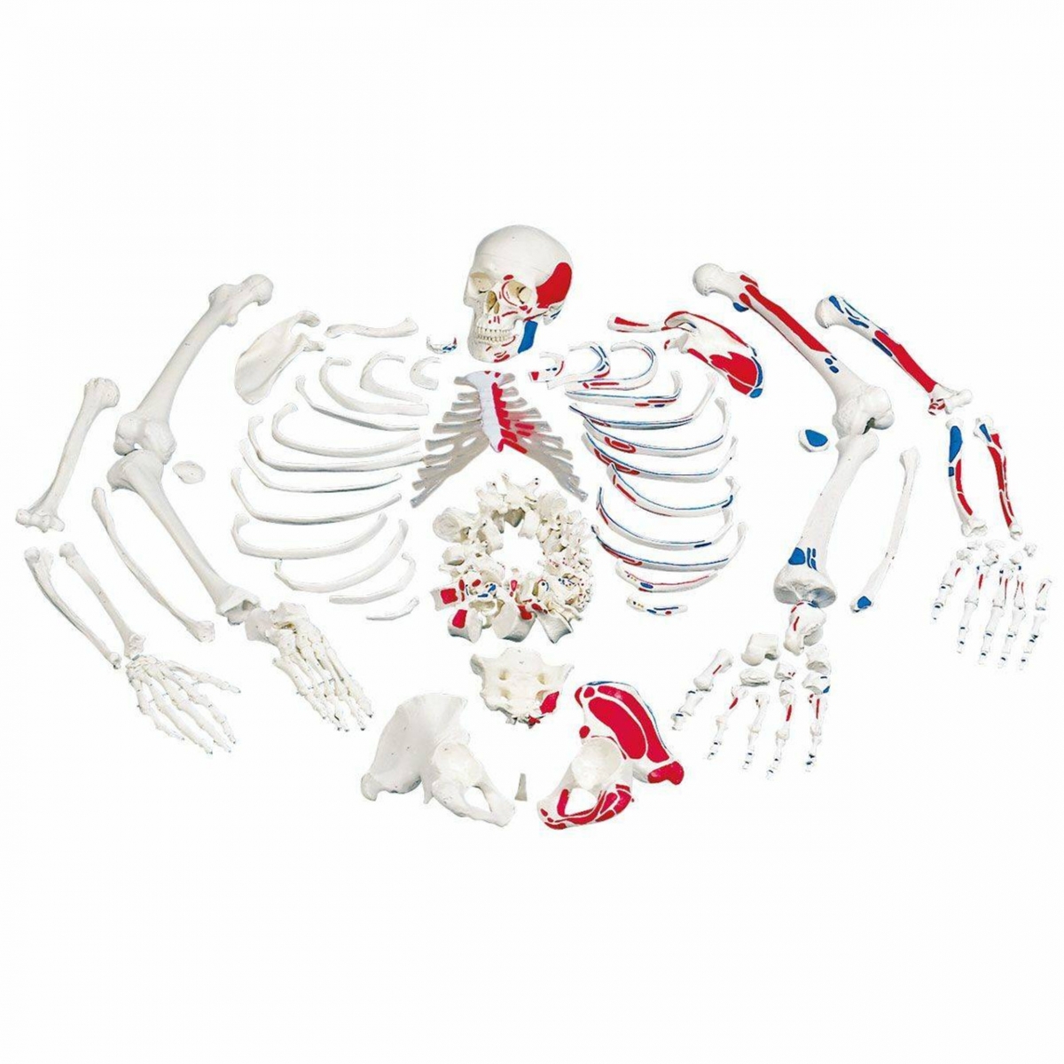
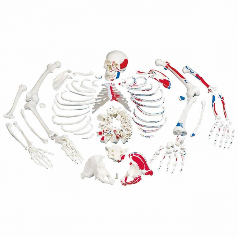
 Bilateral, 96% Anatomical Accuracy, Premium Medical Grade Quality, Complete Disarticulated Human Skeleton Model")
₹ 10,599 ₹ 28,965
 Made of PVC Plastic")
₹ 1,599 ₹ 2,909
 5.8 Feet Tall 96% Anatomical Accuracy Premium Quality | MYASKRO")
₹ 14,699 ₹ 33,039

₹ 1,899 ₹ 5,488

₹ 10,699 ₹ 25,455

₹ 33,599 ₹ 57,779

₹ 19,599 ₹ 25,801

₹ 11,799 ₹ 18,945
The Ultimate Anatomy Study Tool – Now with Muscle Mapping
Bring musculoskeletal anatomy to life with the MYASKRO® Bi-Lateral Disarticulated Human Skeleton Model, meticulously designed for hands-on education in human osteology and muscle function. This advanced teaching tool features a complete set of bones from both sides of the human body, with clearly marked muscle origin and insertion points in red and blue for visual mastery of attachment sites.
Because structure teaches function. This model not only lets you study every individual bone, it also shows where key muscles attach—helping learners understand biomechanics, movement, and muscular coordination. With both sides of the body represented and muscle landmarks highlighted, this is the most comprehensive anatomical bone set in its class.
This full skeletal system boneset includes:
This model supports in-depth learning of:
Perfect for use in anatomy labs, exam prep, or clinical training, this is a must-have tool for anyone studying or teaching musculoskeletal systems at an advanced level.
The MYASKRO® skeleton model is designed for serious academic use. Whether it’s in a classroom, clinical setting, or personal study space, the bones are durable, washable, and packaged to remain organized over time. Each painted landmark aligns with anatomical convention—making the model as informative as it is visually striking.
Yes. All bones are life-sized, modeled to reflect real human dimensions for proper anatomical instruction.
Yes. This is a bi-lateral model, which includes both left and right side bones for complete comparative study.
Red indicates muscle origin points, while blue shows muscle insertion points. These colors follow standard anatomical teaching conventions.
Absolutely. This model is used in medical, physiotherapy, and sports science programs around the world to teach in-depth osteological and muscular anatomy.
You can use removable labels or washable markers for temporary marking. The surface is smooth but durable enough to handle classroom handling and study annotations.
This is more than a bone set—it’s a fully functional teaching platform for serious anatomical exploration.
“We use this model in our orthopedics unit—it’s the best way to teach both skeletal form and muscle function simultaneously.”
– Dr. M. Jadhav, Physiotherapy Professor“Exactly what I needed for my OSCE revision. Having both sides lets me compare symmetry and remember insertions faster.”
– Aarav Mehta, Medical Student“We bought two for our PT school. It’s durable, detailed, and the red-blue muscle points are incredibly helpful for visual learners.”
– Sneha Rao, Academic Lab Technician
Each bone is securely packaged and labeled for easy unpacking and storage. The entire set comes boxed and protected, with bones grouped by region (skull, vertebrae, thorax, limbs, etc.) to save prep time and keep your lab organized.
Study human anatomy from the inside out—accurately, interactively, and in full color. Whether you’re teaching skeletal alignment, tracing muscular attachments, or reviewing joint mechanics, this model offers a hands-on learning experience unmatched by charts or software.