


₹ 8,499 ₹ 22,445
 - Myaskro")
₹ 8,999 ₹ 22,379

₹ 5,799 ₹ 15,157
 5.8 Feet Tall 96% Anatomical Accuracy Premium Quality | MYASKRO")
₹ 14,699 ₹ 33,039

₹ 33,599 ₹ 57,779
")
₹ 43,899 ₹ 58,965
 | Myaskro")
₹ 44,299 ₹ 58,945

₹ 31,699 ₹ 42,645
 | Myaskro")
₹ 3,199 ₹ 4,865
")
₹ 3,199 ₹ 4,895
")
₹ 6,399 ₹ 8,945
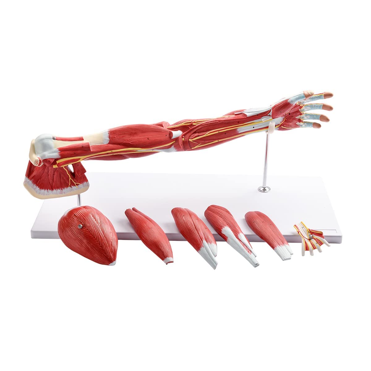
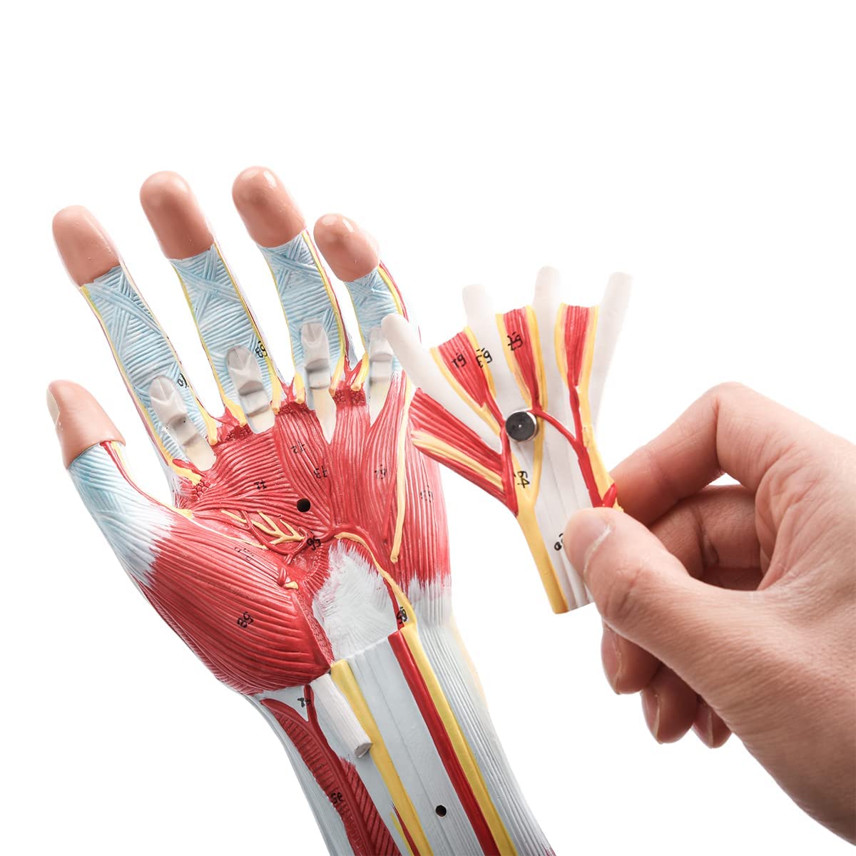
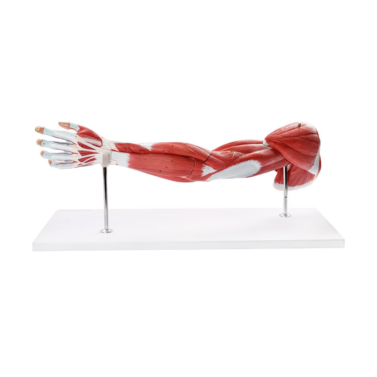
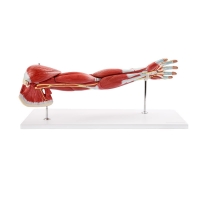
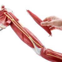
Muscular Human Arm Model:
Detailed anatomical model of the upper limb, demonstrating superficial and deep musculature, tendons, nerves, and vascular pathways.
Includes removable muscle groups: deltoid, biceps brachii, triceps brachii, brachialis, and additional forearm flexor/extensor muscle compartments.
Displays the course of major neurovascular bundles including the brachial artery, median, radial, and ulnar nerves.
Precisely delineates insertion and origin points, facilitating instruction on musculoskeletal biomechanics, innervation, and compartmental anatomy.
Essential for didactic purposes in anatomy, orthopaedics, physical therapy, neurology, and surgical training.
This Muscular Human Arm Anatomical Model offers a comprehensive representation of the musculature, neurovascular structures, and tendon insertions of the upper limb. The model features multiple detachable muscle groups—deltoid, biceps brachii, triceps brachii, brachialis, and select forearm muscle compartments—enabling detailed examination of both superficial and deep layers. Clearly marked origins and insertions allow for understanding of biomechanical actions and functional anatomy.
Neurovascular elements are rendered with high fidelity, demonstrating the trajectories of the brachial artery, median nerve, radial nerve, and ulnar nerve, as well as associated veins and branches. This model is indispensable for instruction in gross anatomy, clinical orthopaedics, physical and occupational therapy, and for surgical planning and procedural demonstration. It enables a hands-on approach to understanding upper limb pathology, compartment syndromes, nerve entrapments, and reconstructive surgical techniques.
Total Reviews (0)