
₹ 1,299 ₹ 5,389
 with Soft Gingiva And Screw Driver - MYASKRO")
₹ 2,599 ₹ 14,095

₹ 299 ₹ 1,115

₹ 3,499 ₹ 6,118

₹ 13,299 ₹ 18,709

₹ 12,399 ₹ 22,379

₹ 9,299 ₹ 22,355
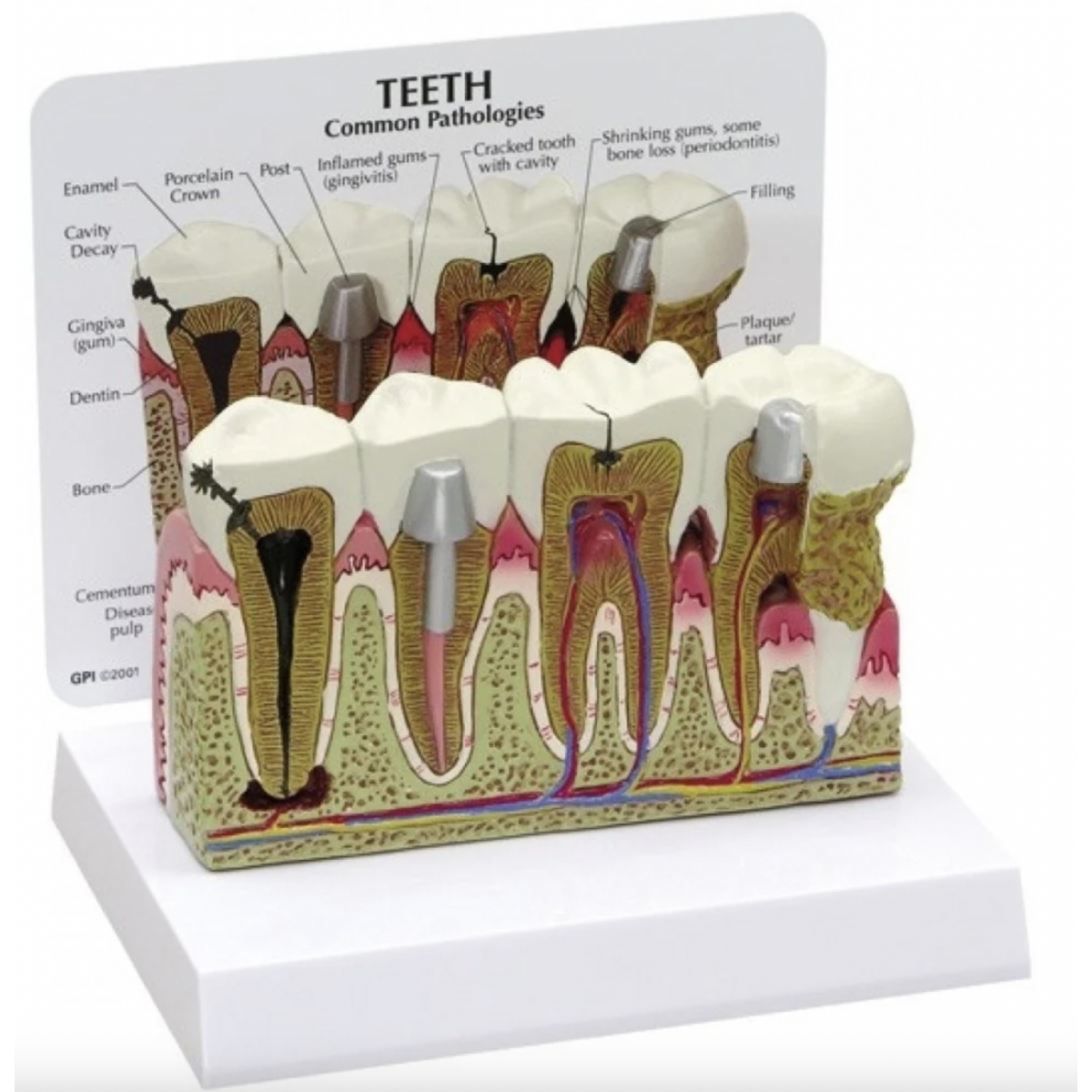
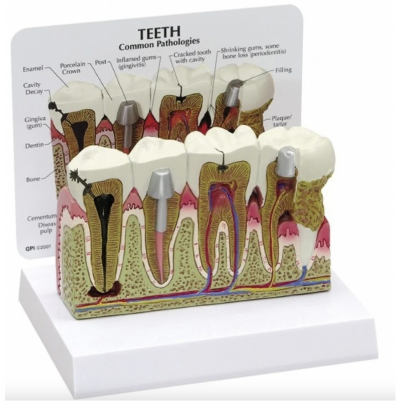
Diseased Teeth and Gums Model – Myaskro
Make pathology visible—from CEJ to apex.
This enlarged multi-tooth cross-section demonstrates the full spectrum of common dental diseases and restorations, helping you convert complex chairside explanations into clear visuals your patients and interns understand.
Caries pathway, end to end: enamel cavitation → dentinal involvement → pulpitis → periapical lesion/abscess—ideal for discussing RCT vs. extraction.
Periodontal disease continuum: plaque/calculus deposition, gingivitis (edematous marginal gingiva), progression to periodontitis with recession, pocketing, and crestal/alveolar bone loss.
Restorative scenarios: amalgam/composite filling, full-coverage crown (porcelain) and post–core presentation for prognosis discussions.
Clear layers for teaching: enamel, dentin, cementum, pulp, PDL region, lamina dura, alveolar bone.
Chairside ready: numbered/labelled legend card; wipe-clean PVC/resin; stable desktop base for OPD, camps, and UG teaching.
Etiology and consequences of untreated caries (reversible vs. irreversible pulpitis, apical pathology).
Biofilm → calculus → gingival inflammation → attachment loss; need for SRP/periodontal surgery and maintenance.
Indications for crowns and post–core after endodontic therapy.
Prevention message: technique, recall interval, and risk-based care.
Total Reviews (0)